
Equipment
The Microscopy and Microanalysis Research Laboratory (MMRL) is a research unit at the College of Science and Mathematics at Montclair State University. The MMRL is University wide resource and facility. Access to the MMRL and its microscopes is open to all potential University users who have a need for the facilities in their research and classes, as well as external public and private sector clients.
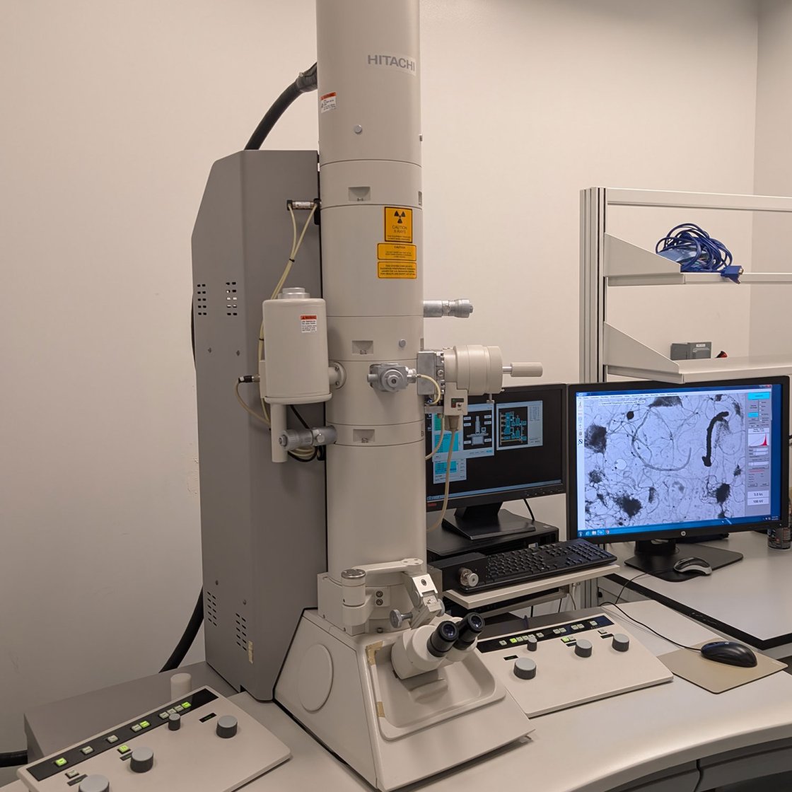
Hitachi H-7500 TEM
The Hitachi H-7500 tungsten/LaB6 TEM is an instrument suitable for high magnification examination of biological tissues and soft materials. It is fitted with Bruker thin-window energy dispersive X-ray (EDS) detector which is capable of detecting elements ranging from boron upwards. A slow scan AMT CCD camera with pixel resolution 1024 x1024 is fitted side mount to the column.
The status of the electron microscope is displayed graphically on TEM computer, which allows the user to call out window for setting parameters to suit their needs.
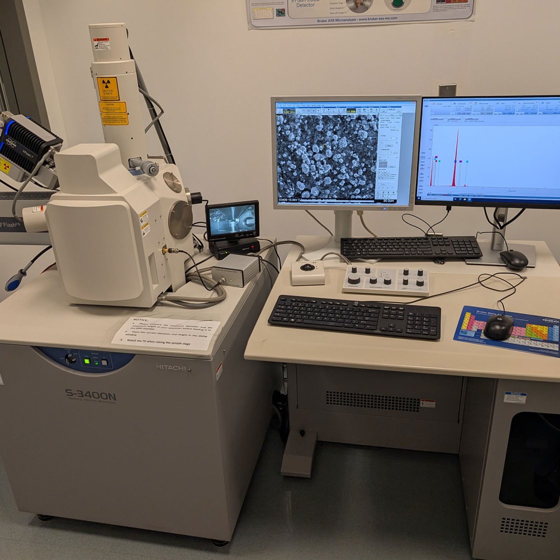
Hitachi S-3400N SEM
The Hitachi S-3400N is a Variable Pressure SEM (VP-SEM) offering advances in automatic l filament saturation and “no touch” objective aperture alignment. High take off angle ports were built to the analytical chamber for detectors such as EDS. A Quad-type semiconductor BSE detector is mounted at the bottom of the objective porepiece, allowing compositional and high resolution imaging at a high or low vacuum mode.
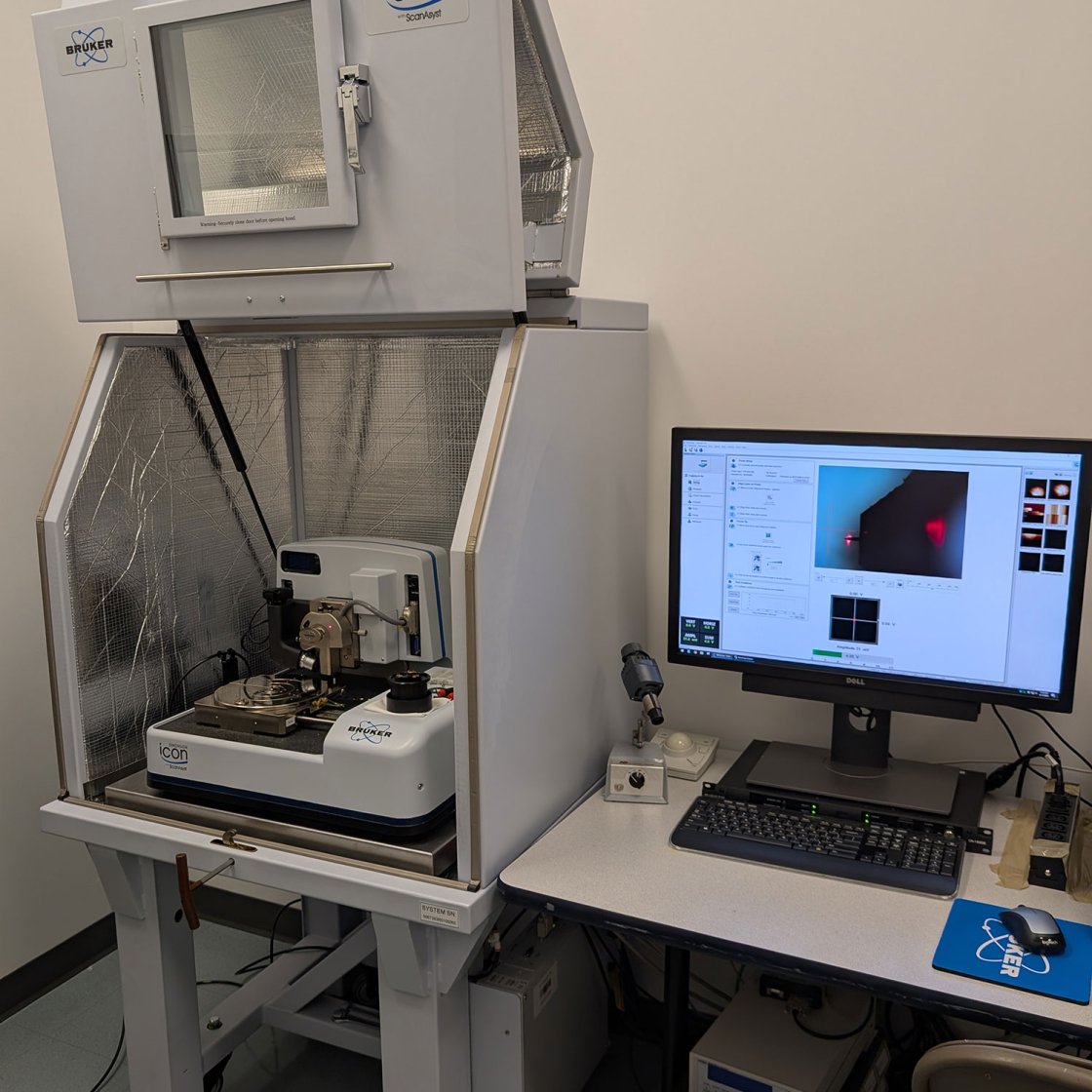
Bruker Dimension ICON® SPM
Bruker Dimension ICON® Scanning Probe Microscope (SPM) brings the highest levels of performance, functionality, and AFM accessibility to nanoscale research. It is equipped with ScanAsyst® automatic image optimization technology based on PeakForce Tapping mode, which performs a very fast force curve at every pixel in the image. This technology allows users to obtain consistent high-quality results faster and easier. The Bruker ICON SPM system is capable of many Imaging Modes and Capabilities.
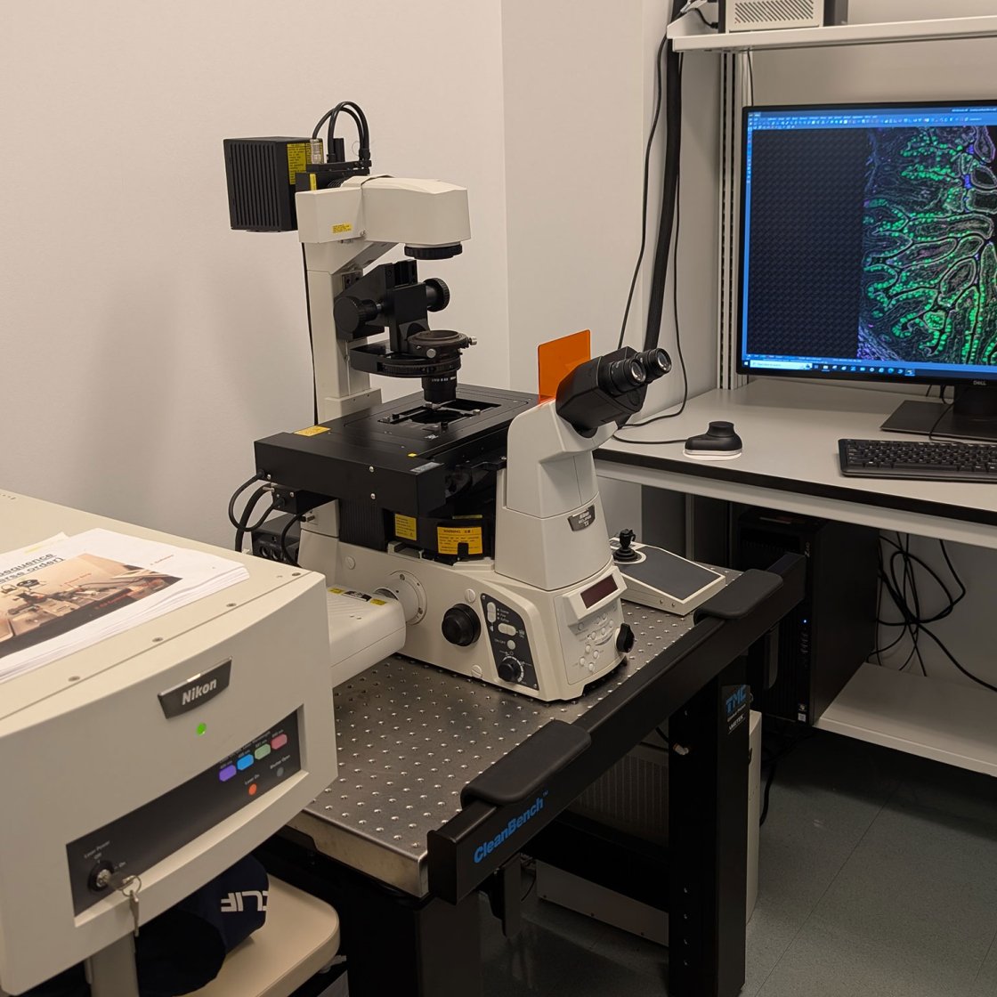
Nikon C2 Plus Confocal Laser Scanning Microscope (CLSM)
The Nikon C2 Plus is a point scanning confocal microscope with four channels: 405, 488, 561 and 640nm excitation. It is equipped with four solid state laser lines (405, 488, 561 and 640 nm) and is able to take images of up to 2048 x 2048 µm pixels resolution. It provides an “X-cite” 120 LED system for fluorescence and filters for widefield using DAPI, GFP, and Tex Red filters. The objectives for the Ti system are 10x, 20x, 40x, 20x oil, 40x oil, 60x oil, and 100 x oil, all with DIC capabilities. Detectors include three standard Photo-multiplier Tubes (PMTs), pco.edge 4.2 scientific CMOS camera, and a transmitted detector. The scan head is attached to a fully automated Nikon Ti microscope and entirely controlled by the NIS-Elements C software. Extended life experiments are facilitated by a Nikon Perfect Focus System that continuously determines the distance to the coverslip.
Other Equipment
Denton Desk IV TSC Cold Sputter/Etch and Carbon Rod Accessory
This tool is used to sputter a conductive film (gold or gold/palladium) on samples for SEM analysis and to provide some limited surface clearing in some models. The mounting of a turbo molecular pump to the system enables it to quickly pump the chamber to a high vacuum level. Six main operations are available for the system: vacuum, etch, rotating, sputter, time etch and time sputter.
Denton DV-502A High Vacuum Evaporator
DV-502A is a high vacuum evaporator for fine carbon (for X-ray microanalysis) or metal evaporation of scanning and transmission electron microscopy samples. Using Denton’s economical diffusion pump design, it can rapidly and repeatedly cycle from atmosphere to high vacuum.
Carbon coatings can be evaporated onto grids as a support layer or as an additional stabilizing agent for polymer coated grids. Grids that will be used for imaging isolated macromolecules (such as proteins) can be glow discharged to decrease their hydrophobicity.
Denton Vacuum DCP-1 Critical Point Dryer
The Denton critical point drying apparatus is mounted on a sturdy stand with a weighted base which sits conveniently on the laboratory bench. It is being used to critical point of liquid carbon dioxide (CO2) to dry biological material with a graded series of ethanol. The chamber is 1-1/8” dia. × 2-5/8” high and will hold a large number of grids or a half dozen SEM stubs. A removable stainless steel wire basket 1” dia. × 2-1/2” long is also supplied.
Leica 2030 rotary paraffin microtome
Reichert/Leica 2030 Microtome is designed for most routine histology and biology needs. High precision and stability ensures reproducible section quality in wax and plastic embedded sections. It has a smooth handwheel movement that allows controllable sectioning speed with zero backlash. Vertical roller veering sideway prevents any deviation of specimen cylinder during sectioning. A section counter and the automatic specimen feed is 1 to 60 um. Three Reichert microtomes are available in our EM facilities.
Leica Reichert Ultratrim
Leica Reichert ultratrim is an EM specimen trimmer built-in high speed milling device. The ultratrim consists of stereo microscope with 20x total magnification, illuminator 20W reflector halogen light, specimen carrier and trimming tool. Trimming is carried out during observation of the specimen with the microscope, using a special milling tool which is moved past the specimen. The specimen can be centered ± 2 mm and rotated about 90°C click stop mechanism for milling the pyramid faces. The selected detail remains in the center of the field during each operation.
Leica EM UC7 Ultramicrotome
The Leica EM UC7 offers advanced ultramicrotome technology and features a touch screen control panel. It provides easy preparation of semi- and ultrathin sections as well as perfect, smooth surfaces of biological and industrial samples for TEM, SEM, AFM and LM examination. Three independent, built-in, brightness-controlled LED light sources and the additional LED spotlights provide outstanding illumination. The innovative touch-sensitive control unit of the Leica EM UC7 enables fast and safe alignment of knife and specimen with help files and prompts to hand for beginners. Programmable knife and cutting movements make trimming easy.
Tissue-TeK VIP Tissue Processor
EM tissue processor provides a compact system for automatically processing biological specimens for high resolution light microscope examination.
Thermo Scientific HistoStar Embedding Workstation
Thermo Scientific HistoStar Embedding Workstation consists basically of a control panel, paraffin storage reservoir and dispenser, embedding module with hot and cold spots for orientation, warm tissue storage drawer, mold storage, cold plate, and a waste drawer for collecting excess paraffin from the working platform. The HistoStar embedding workstation is the only workstation with a heated wax trimmer built directly into the workspace. It is the well-illuminated, ergonomic, efficient workstation.